Miten laparoskooppinen kolekystektomia on
Dec 08, 2021
Laparoskooppisesta kolekystektomiasta on tullut kypsä kirurginen tekniikka, jonka suurin osa potilaista hyväksyy, joille on ominaista vähemmän trauma, vähemmän kipua ja nopea toipuminen.
(1) Merkinnät
① Symptomatic gallstones.
② Symptomatic chronic cholecystitis.
③ Gallstone with diameter >3 cm.
④ Filled gallstones.
⑤ Symptomatic and surgically indicated protuberant lesions of the gallbladder.
⑥ The symptoms of acute cholecystitis were relieved after treatment, and there were surgical indications.
⑦ It is estimated that the patient is well tolerated.
(2) Suhteelliset vasta-aiheet
① Acute attack of calculous cholecystitis.
② Chronic atrophic calculous cholecystitis.
③ Secondary choledocholithiasis.
④ History of upper abdominal surgery.
⑤ Fat body.
⑥ External abdominal hernia.
(3) Absoluuttinen vasta-aihe
① Acute cholecystitis with serious complications, such as gallbladder empyema, gangrene, perforation, etc.
② Gallstone acute pancreatitis.
③ With acute cholangitis.
④ Primary common bile duct stones and intrahepatic bile duct stones.
⑤ Obstructive jaundice.
⑥ Gallbladder cancer.
⑦ Protuberant lesions of the gallbladder are suspected to be cancerous.
⑧ Cirrhosis and portal hypertension.
⑨ Middle and late pregnancy.
⑩ Abdominal infection, peritonitis.
Chronic atrophic cholecystitis, gallbladder less than 4.5cm × 1.5cm, wall thickness >0,5 cm (ultraäänimittaus).
Mukana verenvuototaudit ja hyytymishäiriöt.
Henkilöt, joilla on epätäydellinen tärkeiden elinten toiminta, vaikeasti siedettävä leikkaus ja anestesia sekä ne, joilla on sydämentahdistin (sähkökoagulaatio ja sähkökautisointi ovat kiellettyjä).
Yleiskunto on huono, se ei sovellu leikkaukseen tai potilas on vanha, kolekystektomiasta, palleatyrästä ei ole vahvaa viittausta.
Laparoskooppisen leikkauksen käyttöaiheet laajenevat tekniikan kehittyessä. Joitakin sairauksia, jotka olivat alun perin vasta-aiheita leikkaukselle, on myös yritetty saada loppuun laparoskopialla. Jos sekundaarinen koledokolitiaasi on osittain ratkaistu laparoskooppisella leikkauksella. Tarvittavan kokemuksen hankkimisen jälkeen lisää sairauksia voidaan hoitaa laparoskooppisella leikkauksella.
(4) Kirurginen toimenpide
① Create pneumoperitoneum. Make an arc incision along the lower edge of the umbilical fossa, about 10mm long. If the lower abdomen has been operated on, cut the skin on the upper edge of the umbilical fossa to avoid the original surgical scar.
Operaattori ja ensimmäinen avustaja pitävät kumpikin kangaspyyhepihdeistä kiinni vatsan seinämän nostamiseksi napakuopan molemmilta puolilta. Operaattori piti pneumoperitoneum-neulasta (Veress-neula) oikean kätensä peukalolla ja etusormella, kohdistai voimaa ranteeseensa ja puukotti vatsaonteloon pystysuoraan tai hieman vinosti lantiononteloon.
Punktioprosessissa, kun neula murtuu faskian ja vatsakalvon läpi, läpimurto tuntuu kahdesti; Arvioi, onko neulan kärki mennyt vatsaonteloon. Normaalia suolaliuosta sisältävä ruisku voidaan liittää. Kun neulan kärki on vatsaontelossa, se osoittaa negatiivista painetta. Liitä pneumoperitoneum-kone. Jos täyttöpaine ei ylitä 1,73 kpa, se tarkoittaa, että pneumoperitoneumin neula on vatsaontelossa. Älä täytä liian nopeasti alussa. Käytä alhaista täyttöä, 1 2L minuutissa.
At the same time, observe the intraperitoneal pressure on the pneumoperitoneum machine. The pressure during inflation should not exceed 1.73kpa. If it is too high, it indicates that the position of the pneumoperitoneum needle is incorrect, the anesthesia is too shallow and the muscle is not loose enough. Appropriate adjustment should be made. When the abdomen begins to bulge and the liver dullness boundary disappears, it can be changed to high flow automatic inflation until the predetermined value (1.73 2.00kpa) is reached. At this time, the inflation is 3 4L, the patient's abdomen is completely bulged, and the operation can be started.
Lift the abdominal wall with towel pliers at the umbilical pneumoperitoneum needle and puncture with 10mm trocar. The first puncture has a certain "blindness", which is a more dangerous step in laparoscopy. Be extra careful. Rotate the trocar slowly and enter the needle evenly. When entering the abdominal cavity, there is a feeling that the resistance disappears suddenly. Open the closed air valve and gas escapes. This is the success of puncture. Connect the pneumoperitoneum machine to maintain constant pressure in the abdominal cavity. Then put the laparoscope in and puncture at each point under the monitoring of the laparoscope.
Yleensä puhkaise 2 cm xiphoid-prosessin alapuolelle ja aseta 10 mm:n kotelo poistokoukkua, puristinapplikaattoria ja muita instrumentteja varten; Punktio 2 cm oikean keskimmäisen solisluun linjan kylkireunan alapuolelle tai 2 cm suoran vatsalihaksen ulkoreunan ja kainalon etuosan kylkireunan alapuolelle 5 mm:n troakaarilla asettaaksesi kastelulaitteen ja sappirakon kiinteät tartuntapihdit. Tällä hetkellä keinotekoinen pneumoperitoneum ja valmistelut on saatu päätökseen.
Pneumoperitoneumin valmistuksen ja ensimmäisen troakaaripunktion vuoksi vatsaontelon suuret verisuonet ja suolet voivat vahingossa vammautua, eikä niitä ole helppo löytää leikkauksen aikana. Viime aikoina monet ihmiset ovat tehneet napaan pienen aukon löytääkseen vatsakalvon ja laittaneet troakaarin suoraan vatsaonteloon täyttöä varten. Pneumoperitoneumin onnistuneen valmistuksen jälkeen leikkaus aloitettiin.
② Dissect the Calot triangle. Grasp the neck of gallbladder or Hartmann's bursa with grasping forceps and traction to the upper right. It is best to draw the cystic duct perpendicular to the common bile duct in order to clearly distinguish the two, but pay attention not to draw the common bile duct into an angle. The serous membrane on the cystic duct was cut with an electrocoagulation hook, the cystic duct and cystic artery were passively separated, and the common bile duct and common hepatic duct were distinguished. Since it is close to the common bile duct, electrocoagulation should be used as little as possible to avoid accidental injury to the common bile duct. Use the electrocoagulation hook to separate the cystic duct upstream and downstream, and see the relationship between the cystic duct and the common bile duct. Place the titanium clip as close to the gallbladder neck as possible. There should be sufficient distance between the two titanium clips. The titanium clip should be at least 0.5cm away from the common bile duct. Cut between the two titanium clips with scissors, and do not use electric cutting or electrocoagulation to prevent damage to the common bile duct due to heat conduction. Then find the cystic artery behind it and cut it with titanium clip. After cutting off the gallbladder artery, do not pull hard to avoid breaking the gallbladder artery, and pay attention to the posterior branch of the gallbladder. Carefully peel off the gallbladder, electrocoagulation or hemostasis with titanium clip.
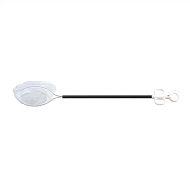
③ Cholecystectomy. Clamp the gallbladder neck and pull it upward, carefully peel it off along the gallbladder wall, and the assistant should assist in pulling to make the gallbladder and liver bed have a certain tension. Completely peel off the gallbladder and place it on the upper right side of the liver. The liver bed was hemostatic by electrocoagulation, carefully rinsed with normal saline, and checked for bleeding and bile leakage (a piece of gauze was disposed at the hepatic hilum, and checked for bile staining after removal). After absorbing all the water in the abdominal cavity, transfer the laparoscope to the lower sleeve of the xiphoid process and give way to the umbilical incision, so that the gallbladder containing stones greater than 1cm can be taken out from the umbilical incision with loose structure and easy expansion. If the stones are small, they can also be taken out from the puncture hole under the xiphoid process.
④ Remove the gallbladder. Put the toothed claw forceps into the abdominal cavity from the cannula at the umbilicus, grasp the residual end of the cystic duct under monitoring, slowly drag the gallbladder into the cannula sheath and pull it out together with the cannula sheath. When grasping the gallbladder, pay attention to placing the gallbladder on the liver to avoid accidental injury to the intestinal canal by sharp forceps. If the stone is large or the tension of the gallbladder is high, do not pull it out with force to avoid rupture of the gallbladder and leakage of stones and bile into the abdominal cavity. At this time, the incision can be enlarged with vascular forceps and taken out, or the incision can be expanded to 2.0cm with an expander. If the stone is too large, the incision can be extended. If bile leaks into the abdominal cavity, wet gauze shall be used to enter from the umbilical incision to suck up the bile.
Jos kivi on liian iso poistettavaksi viillosta, voit myös avata sappirakon ensin, imeä sappirakosta imuputkella ja ottaa sen yksitellen ulos pihdeillä murskattuaan. Jos huomaat, että kivi putoaa vatsaonteloon, ota se pois. Kun olet tarkistanut, ettei vatsaontelossa ole verta ja nestettä, vedä laparoskooppi ulos, avaa kanyylin venttiili hiilidioksidikaasun poistamiseksi vatsaontelosta ja vedä sitten kanyyli ulos. Viilto 10 mm:n kanyylillä ommellaan ohuella langalla fasciakerroksena 1 2 pistoa varten ja jokainen viilto suljetaan steriilillä liimakalvolla.
(5) Suuret komplikaatiot
① Bile duct injury. Bile duct injury is one of the most common and serious complications of laparoscopic cholecystectomy.
Sappitievaurioiden ja sappivuotojen ilmaantuvuus on noin 10 prosenttia. Siihen tulee kiinnittää riittävästi huomiota. Se johtuu pääasiassa Calot-kolmion epäselvästä anatomiasta, erityisesti valppauden puutteesta yhteisen sappitiehyen tai kystisen tiehyen yleistä vaihtelua vastaan. Kystistä tiehaa erotettaessa sappitiehy vaurioitui vahingossa lämpövaurioon, sappivuotoa ei leikkauksen aikana esiintynyt, ja nekroosi ja kudoksen irtoaminen lämpövaurioituneella alueella leikkauksen jälkeen saattoi myös aiheuttaa sappivuotoa. Lisäksi sappirakon sängyssä on usein suuria vagaalisia sappitiehyitä. Leikkauksensisäinen sähkökoagulaatio ei voi koaguloitua täysin, ja myös sappivuoto voi muodostua. Tärkeimmät sappitievaurioiden ilmenemismuodot ovat voimakas ylävatsakipu, korkea kuume ja keltaisuus. Potilaat, joilla on tyypillisiä oireita, hoidetaan yleensä ajoissa leikkauksen jälkeen; Muutamilla potilailla esiintyi kuitenkin vain vatsan turvotusta, ruokahaluttomuutta, alhaista kuumetta ja etenevää pahenemista. Tällaisia potilaita tulee tarkkailla tarkasti. Raportin mukaan vatsansisäistä sapen kertymistä havaittiin muutama kuukausi leikkauksen jälkeen. Sappivuodon arvioiminen riippuu pääasiassa ultraäänestä tai TT:stä, ja sen jälkeen varmistetaan hienolla neulapistoksella ultraäänen tai CT:n tai radionuklidihepatokolangiografian ohjauksessa.
② Vascular injury. One is massive hemorrhage caused by needle tip injury to abdominal aorta, iliac artery or mesenteric vessels during pneumoperitoneum and trocar placement. There are many reports of death caused by trocar puncture. Therefore, after successful pneumoperitoneum, laparoscopy should peep the whole abdomen once to prevent missing vascular injury.
Toinen on maksaportin epäselvä anatomia tai oikean maksavaltimon tai oikean maksavaltimon väärä kiinnitys sappirakon valtimon verenvuodon vuoksi. On myös raportoitu porttilaskimovaurioita anatomian aikana. On raportoitu oikeanpuoleisen maksan nekroosia, joka johtuu maksavaltimon väärästä puristamisesta.
③ Intestinal injury. Intestinal injuries are mostly accidental injuries caused by electrocoagulation, mainly because the electrocoagulation hook is not placed in the TV monitoring picture and is not found. Abdominal pain, abdominal distention and fever occur after operation, resulting in serious peritonitis, and its mortality is high.
④ Postoperative intraperitoneal hemorrhage. Postoperative intraperitoneal hemorrhage is also one of the serious complications of laparoscopic surgery. The injured parts are mainly the blood vessels near the gallbladder, such as hepatic artery, portal vein and abdominal aorta or vena cava during periumbilical puncture. The manifestations were hemorrhagic shock, abdominal bulge and peripheral circulatory failure. Open surgery should be performed immediately to stop bleeding.
⑤ Subcutaneous emphysema. The causes of subcutaneous emphysema are as follows: first, when making pneumoperitoneum, the pneumoperitoneum needle did not penetrate the abdominal wall, and high-pressure carbon dioxide entered the subcutaneous; Second, due to the small skin incision, the trocar is embedded very tightly, and the puncture hole of the peritoneum is relatively loose. During the operation, carbon dioxide gas leaks into the lower skin layer of the abdominal wall. Postoperative examination can find abdominal subcutaneous twisting pronunciation, generally without special treatment.
⑥ Others. Such as incisional hernia, incisional infection and abdominal abscess.